Image Processing and Guidance, PC1-PC3
Improve Your Imaging
Tips & tricks to get the best images
- 10 tips for image acquisition
- Image acquisition video tutorials
- Get help with bioimage analysis; DBI-INFRA; (pay for service)
- Guidelines for image acquisition ethics
- Digital imaging ethics: H Pearson, CSI: cell biology; Nature 434, 952-953 (2005)
- CJ Waters, Accuracy and precision in quantitative fluorescence microscopy; J Cell Biol (2009) PMID 19564400
- AJ North, Seeing is believing? A beginners' guide to practical pitfalls in image acquisition; J Cell Biol (2006) PMID: 16390995
- JS Aron et al, Image co-localization - co-occurrence versus correlation, J Cell Sci (2018) PMID: 29439158
- JM Heddleston et al. A guide to accurate reporting in digital image acquisition - can anyone replicate your microscopy data? J Cell Sci (2021) PMID: 33785608
Find a suitable protocol
... or share your protocol and help your colleagues!
Please send us your protocol (Word, pdf) and we will upload it here.
Learn More
Dive deeper into the world of microscopy
A zoom-in on cameras & detectors
For basic and advanced imaging topics
Go Further
Extend your analyses
... with histology
For assistance and guidance on histology matters, contact Helene M. Andersen at the Histology Core Facility located at Biomedicine in the Skou building, 1116, room 270D.
... with Flow Cytometry
For studying host-pathogen interactions, use Imaging Flow Cytometry at the FACS Core Facility at Biomedicine.
Networks & other imaging core facilities
Learn more about national and international imaging infrastructures.
Image Processing PCs
- Registered users may perform advanced image processing using our dedicated Image Processing PCs (PC1-PC3 and HIVE). Please contact The Bioimaging Team to get started.
- Image processing PCs cannot be accessed remotely at the moment.

Open office
Come to our Open Office sessions to discuss your imaging needs or get advice on workflows, free of charge:
Tuesdays and Thursdays
13:30 -14:30
The Skou building, 1116-256
Or contact us on:
- imaging@biomed.au.dk
- or via the contact form below
PC1
3D and 4D image processing and deconvolution
Location: building 1116, room 270E
- Imaris 8.2 single full for 3D/4D visualization and image processing *)
- NOTE: Imaris does not work via Remote Desktop
- imaris converter and viewer freeware
- cellSens dimention v2.3 with Count and Measure and Constrained Iterative Deconvolution
- Scan^R for high content imaging data analysis
- MATLAB
- Freeware Fiji and Ilastik
*) This PC has a Nvidia RTX 2070 graphics card for high performance image processing.
What is deconvolution?
Deconvolution makes your images nice and crisp. This image processing technique improves contrast and resolution of images captured by light microscopy. Out of focus light causes blur which mathematically is represented as a convolution operation. Deconvolution seeks to reassign out of focus light to the correct origin. Nearly all fluorescence images can be deconvolved.
PC2
Zen, cellSens and DIC image acqusition
Location: building 1242, room 156
- Advanced processing with Zeiss Zen Desk software (ei. Airyscan processing)*)
- Simple file processing and conversion of files with cellSens Entry 2.3, i.e. convert and process vsi.-files from the slide scanner
- Acquire DIC images with the inverted bright-field microscope
*) This PC has an ASUS DUAL-GTX1660S-6G-EVO graphics card for high performance image processing.
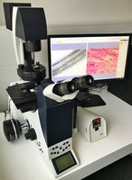
Bright-field DIC microscope

For rutine check of stained and unstained cells or tissue.
Semi-motorized bright-field inverted microscope (no fluorescence) with a sensitive color camera and heating stage, controlled by Image processing PC2.
Objectives Please note: all objectives are dry objectives 5x, 10x (Ph), 20x (Ph), 40x (Ph) 40x (DIC), 63x (DIC) Contrast methods Phase contrast (Ph) and Differential Interference Contrast (DIC) |
PC3
Arivis vision 4D
Location: building 1116, room 550
- Arivis Vision 4D for 2D, 3D and 4D visualization, annotation, and image batch processing regardless of image size
- MATLAB
- Freeware like Fiji and Ilastik
This PC has a Nvidia RTX 2070 super graphics card for high performance image processing.
Software Packages and Freeware
Fiji/ImageJ
Fiji is ImageJ with additional plugins e.g. allowing opening of multiple file formats.
How do I download Fiji/ImageJ?
Fiji can be downleade here.
1. Download the correct version for your system (Win64/32, Mac, Linux64/32)
2. Place the file on your desktop or somewhere else that you administrate ("program files" folder is not recommended)
3. Run the ".exe file" (eg. ImageJ-win64)
4. Accept Update of ImageJ upon first start up
How do I open Olympus (.vsi) files in Fiji?
Go to: https://imagej.nih.gov/ij/docs/guide/146.html
In order to open Olympus (.vsi) files, please follow this guide:
- download Bioformat plugin
- open Fiji folder in file explorer, e.g.: C:\Users\xxxxxx\Documents\Fiji\Fiji.app\plugins
- replace the "Bioformat plugin x.x" with the one you have downloaded and unzipped
- open Fiji - Plugin - Bio-format importer and select your file(s)
QuPath
QuPath is a powerful user-friendly bioimage analysis software designed to extract quantitative information from microscopy images. QuPath is optimised to process whole slide images from digital pathology, but it can also handle 2D images acquired by most fluorescence microscopes.
Common uses include identification, measurement and classification of nuclei, cells and regions of interest in the images.
Cell Profiler/Cell Analyst
Cell Profiler/Cell Analyst – Image Batch Processing and Data Analysis
The scikit-image SciKit (toolkit for SciPy)
The scikit-image SciKit - (toolkit for SciPy) extends scipy.ndimage to provide a versatile set of image processing routines. It is written in the Python language.
ICY
ICY - An open community platform for bioimage informatics
MATLAB
What is MATLAB?
Our sensitive high-resolution microscopes generate large volumes of data that may contain insights linking structure and function at the molecular, cellular, or tissue level, and may be used to decipher mechanisms and energetics of biological processes. The powerful programming software MATLAB may e.g. be used for:
- developing new image processing, statistics, or machine learning techniques not available in commercial software and not packaged with instruments
- automating tedious or compute-intensive workflows such as preprocessing, categorizing, and analyzing collections of images or videos, including collections that are too large to fit into memory
- leveraging available compute resources including multicore computers and GPUs, HPC systems, and the cloud
- deploying analytics and algorithms to large groups of users or collaborators via the web or standalone software
MATLAB is installed on our Image Processing PC1 and PC2 running smoothly thanks to the powerful GPUs of our PCs.
Ilastik
Leverage machine learning algorithms to easily segment, classify, track and count your cells or other experimental data. Most operations are interactive, even on large datasets. You only need to draw the labels and will immediately see the result.
No machine learning expertise is required.
Insight toolkit (ITK)
Insight toolkit (ITK) - an open-source, cross-platform library that provides developers with an extensive suite of software tools for image analysis
The Visualization Toolkit (VTK)
The Visualization Toolkit (VTK) - an open-source software system for 3D computer graphics, modeling, image processing, volume rendering, scientific visualization, and 2D plotting
OpenCV
OpenCV - an open source computer vision and machine learning software library.