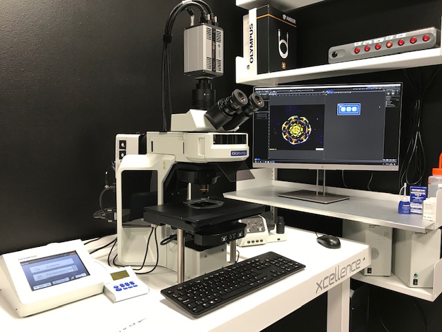
BX63, Upright Widefield, Olympus
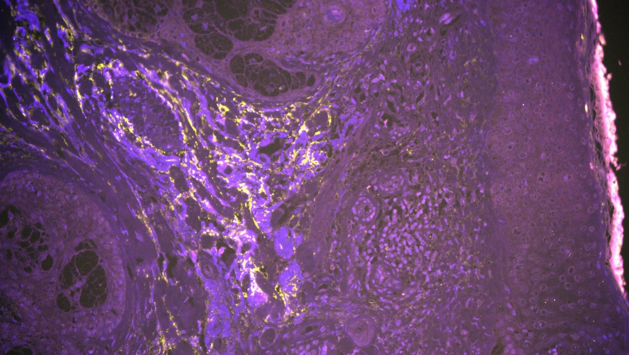
Human skin, Emilia Holm
Would you like your image to be displayed here?
Please contact us or send an email to imaging@biomed.au.dk, if you would like to share your images - Thank you
The BX63 microscope is fully automated and easy to use while maintaining sensitivity.
Applications in brief
The motorized nosepiece automatically focuses onto the sample. The equally motorized stage can either be positioned by hand, enabling rapid gross sample alignment, or it can be encoded to continuously read-out the X and Y position, enabling the user to set precise coordinates and to navigate directly to them.
Features
- Detection of multispectral fluorescence
- Encoding of X and Y coordinates for continuous automated localization
Which samples may I use?
Selected samples, but not limited to:
- fixed tissue sections
- fixed cells
Training and use
Training and use
Tips and tricks
My section has varying thickness or is mounted unevenly. How do I get a crisp image?
- Add multiple focus points
- Go to 'Stage Navigator'
- Choose 'High Density Focus Map'; you will see 5 preselected boxes/focus points
- Press the green arrow button to go through the focus points while adjusting each one
- If needed, add more focus points by right-clicking on the overview and choosing 'Add focus point'
- When all focus points are adjusted, start experiment
Please check all z-heights of focus points before starting the acquisition --> danger of damaging objectives if not correctly set up
Specifications
Specifications
| Full equipment name | BX63 Upright Widefield Fluorescence Microscope, Olympus |
| Camera | Sensitive Andor Zyla 5.5 (5 Megapixel, 6.5 µm pixel – 60% Quantum efficiency) |
| Objectives |
|
| Filter qubes |
|
| Light source |
|