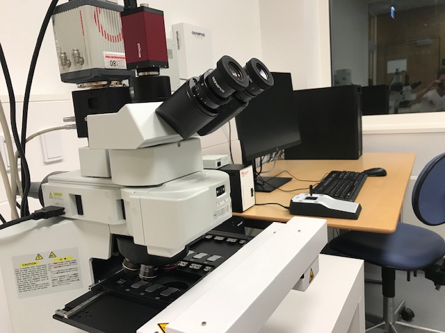
Slide Scanner, Upright Widefield, Olympus


Would you like your image to be displayed here?


Please contact us or send an email to imaging@biomed.au.dk, if you would like to share your images - Thank you


The VS120 Slide Scanner from Olympus is convenient for automated imaging of tissue sections located on several slides.

Applications in brief

This virtual 6-slide scanning system creates high resolution images in brightfield (all magnifications), darkfield (4x, 10x, and 20x) and / or multi-fluorescence mode (all magnifications). Image stitching is very precise enabling high-level accuracy that can be applied from small animal brain slices to large specimens. Seamless switching between micro and macro observation modes enable swift viewing of regions of interest and overall structures.  

Features

  • Brightfield, darkfield, and widefield fluorescence scanning
  • Color corrected camera for brightfield/darkfield scanning
  • Darkfield mode to increase contrast in unstained samples 
  • Fast and powerful light engine with single-band exiters and a fast filter wheel with single-band emitters for DAPI, FITC, Cy3, Cy5 and Cy7

Which samples may I use?

Selected samples, but not limited to:

  • tissue sections
  • up to 6 slides simultaneously

Specifications

Full equipment name VS120 Slide Scanner, Olympus
Objectives
  • 2x / NA 0.08
  • 10x / NA 0.4
  • 20x / NA 0.75
  • 40x / NA 0.95
Channels
  • DAPI: EX 395/25; 295mW, EM 425/50
  • FITC: EX475/28; 196mW, EM 525/50
  • Cy3: EX 575/25; 310mW, EM 600/60
  • Cy5: EX 635/22; 231mW, EM 680/80
  • Cy7: EX 730/40, EM 800/100
Filter DAPI/FITC/Cy3/Cy5/Cy7 Pentafilter Semrock, Penta LED HC Filter Set #F68-050) 
Cameras
  • Monochrome camera: Hamamatsu ORCA-FLASH4.0 V2 (QE 82%)
  • Color camera: AVT Pike F-505C VC50 progressive scan CCD camera
Slide capacity up to 6 slides simultaneously

FAQ

Equipment

- How should I clean the equipment after use?

  • Please leave the work station neat and clean for the next user.

- Are consumables included in the price?

  • Experimental consumables are not included in the price for booking of microscopes via the Bioimaging Core Facility.
  • Designated oil for the oil objectives can be found next to the microscope. Please only use this designated oil for the objectives.
  • Lens paper designated for cleaning of objects as well as designated tissues for cleaning of samples are located next to the microscope.

Data Storage

- How can I get access to my data?

  • After using a microscope or a workstation, please transfer your data immediately to your personal AU drive (O:) and clean up your local directories on instrument PCs if you are not reusing data in subsequent experiments.
  • The Bioimaging Core Facility staff will provide support to non-AU users for exporting data.
  • Use of USB memory sticks or external hard drives is for security reasons not allowed.

- Does the Bioimaging Core Facility regularly back up the user data?

The Bioimaging Core Faility is not responsible for your data. DATA ON LOCAL PC DRIVES WILL BE DELETED ON A REGULAR BASIS in order to keep the systems performing optimally.

Please ensure that you transfer your data to your personal AU drive (O:) after each session.

- May I use my personal USB memory stick or external hard drive for data storage?

No, any use of USB memory sticks or external hard drives is for security reasons not allowed. Usage of USB memory sticks may lead to suspension of the user license.

Data Analysis

- How may I process and analyse my data?

  • Obtained images may be processed and analyzed using freeware or a selection of software available on PCs in the Bioimaging Core Facility.

Publications

- How shall I refer to the equipment?

Need help?

Come to our Open Office sessions to discuss your imaging needs or get advice on workflows, free of charge:

Tuesdays and Thursdays 13:30 -14:30

The Skou building, 1116-256