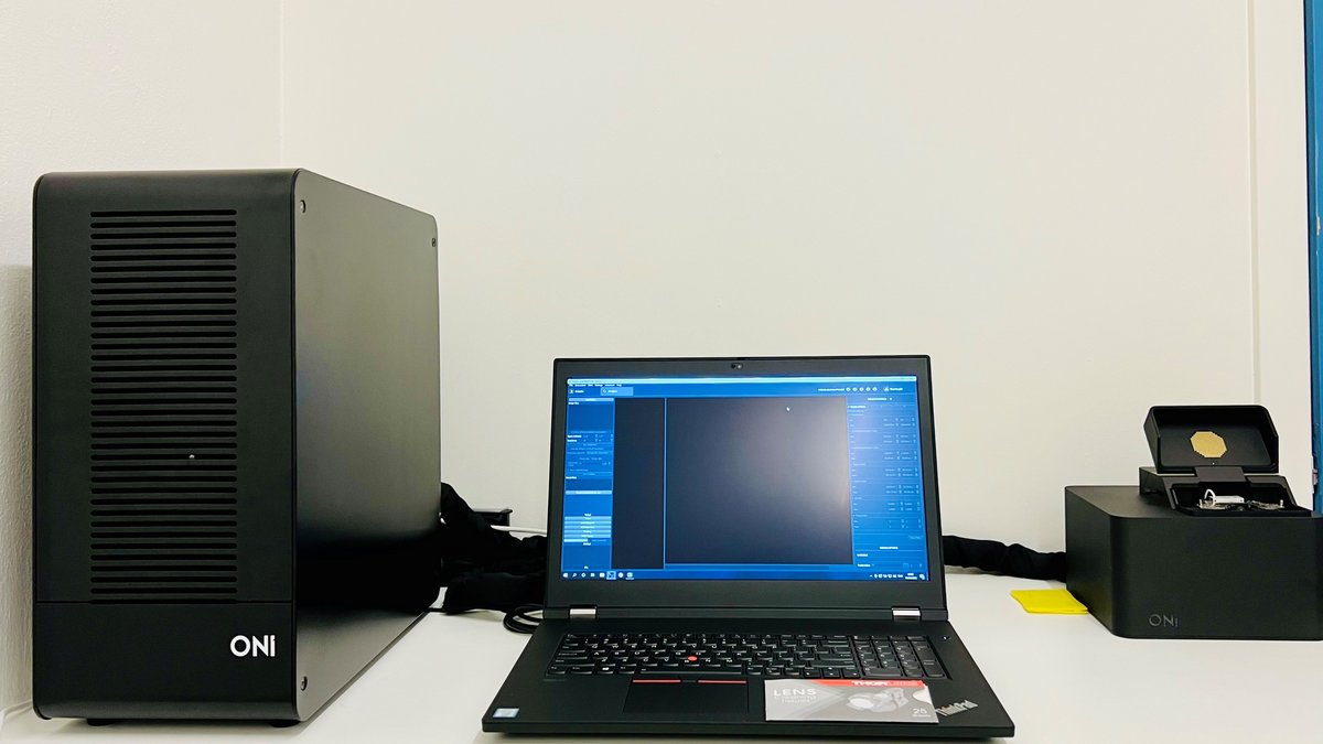
Nanoimager, ONI


Would you like your image to be displayed here?


Please contact us or send an email to imaging@biomed.au.dk, if you would like to share your images - Thank you


The Nanoimager super-resolution microscope from ONI allows investigations down to single-molecule level.

Applications in brief

With its super-resolution capacity, this microscope is excellent for analyses down to single-molecule level, e.g. allowing

  • observation and tracking of single molecules, i.e. spatial distribution
  • biomarker quantification
  • nanoscale morphology
  • investigations of molecular interactions in intracellular pathways
  • localization of extracellular vesicles for cell-to-cell communication
  • characterization of pathogens (ensure that you comply with local safety regulations)

Features

  • Resolution down to 20 nm
  • Single-molecule localization in multiple colors and 2 channels
  • Live-cell imaging facilitating single-particle tracking
  • Precise control and measurement of temperature with heating elements keeping the entire microscope unit at a stable, uniform temperature
  • Up to 1W laser power at the source; 8-12 kW/cm² power density at the sample plane
  • Rapid and precise stabilization of microscope focus with locking technology to minimize Z-drift
  • Post-acquisition drift correction with nanometer precision

Which samples may I use?

Selected samples, but not limited to:

  • tissue sections
  • fixed cells
  • live cells

Specifications

Full equipment name Nanoimager, ONI
Objective Standard, Olympus UPLXAPO100       
Mag./NA 100x/1.45, WD 0.13 mm
Medium Oil
Lasers                                               Type and Power   Selected dyes
405 nm Diode, 1 W DAPI, Hoechst 33342
488 nm Diode, 1 W Alexa488, Atto488, CF488A
561 nm DPSS, 500 mW Alexa555, Dylight555, CF568
640 nm Diode, 1 W Alexa647
Total number of imaging colors Simultaneous imaging channels
  • Up to 4 lasers
  • 1 or 2
Camera
  • Latest generation sCMOS 82 % peak QE, 1.6 electrons rms read noise at standard scan
  • Magnification: 1 pixel = 117 nm in the sample plane
  • Acquisition speed: 100 fps full frame; 5 kHz with frame height cropped to 2%
sCMOS camera
  • Hamamatsu ORCA-Flash4.0 V3 Digital CMOS camera
Illumination modes
  • Closed-loop, continuous illumination angle adjustment between epi-illumination and total-internal reflection
Imaging Modalities
  • Single-molecule imaging
  • 3D localization microscopy
  • Förster resonance energy transfer (FRET) spectroscopy
Field of view
  • 100 μm x 100 μm (1 channel) or 100 μm x 50 μm (2 channels)
Achievable Resolution
  • Lateral: better than 20 nm
  • Axial: better than 50 nm
Other light sources
  • LED for bright-field imaging, NIR auto-focus laser
3D imaging technique
  • Astigmatism
Emission Channels
  • Green (channel 0): band 1: 498-551 nm; band 2: 576-620nm
  • Red (channel 1): 665-705 nm
Focus system
  • One-shot autofocus; Continuous autofocus
Time for super-resolution full frame
  • Seconds to minutes (number of localizations and laser power dependent)
Sample stage
  • 18/18/9 mm XYZ travel range, closed-loop piezo stage with 2 nm encoder resolution
Mechanical stability
  • <1 μm/K drift; <1 nm vibration amplitude (1 Hz to 500 Hz)
Temperature control (live cell imaging)
  • Resistive heating, whole instrument

Special Features

  • Bench-top device; class 1 laser product
  • Homogeneous illumination
  • <10 nm channel mapping accuracy
  • Sensor array (temperature, humidity, CO2, acceleration)
  • Closed-loop adjustments of laser power density at sample plane

Software Features

  • Real-time localization analysis and rendering (sCMOS optimized)
  • Real-time FRET trace analysis
  • Automated 3D sample scanning and acquisition
  • Clustering and co-localization analysis
  • Residual drift correction
  • Scripting interface; OMERO compatibility

FAQ

Equipment

- Which slides and chambers are compatible with the ONI Nanoimager?

Coverslips: optimally 1.5H coverslips, which have 170 µm thickness

Chambers: optimally with #1.5 glass bottom

  • Sample chambers/coverslips must be less than 97 mm long, 35 mm wide and 13 mm in height 
- Are consumables included in the price?

  • No, consumables such as plates and coverslips are not included in the price for booking of the ONI Nanoimager via the Bioimaging Core Facility.

Data Storage

- How can I get access to my data?

  • After using a microscope or a workstation, please transfer your data immediately to your personal AU drive (O:) and clean up your local directories on instrument PCs if you are not reusing data in subsequent experiments.
  • The Bioimaging Core Facility staff will provide support to non-AU users for exporting data.
  • Use of USB memory sticks or external hard drives is for security reasons not allowed.

- Does the Bioimaging Core Facility regularly back up the user data?

The Bioimaging Core Faility is not responsible for your data. DATA ON LOCAL PC DRIVES WILL BE DELETED ON A REGULAR BASIS in order to keep the systems performing optimally.

Please ensure that you transfer your data to your personal AU drive (O:) after each session.

- May I use my personal USB memory stick or external hard drive for data storage?

No, any use of USB memory sticks or external hard drives is for security reasons not allowed. Usage of USB memory sticks may lead to suspension of the user license.

Data Analysis

- How may I process and analyse my data?

  • Data can be exported in standard file formats (*.tiff and *.csv).
  • Currently, the Nanoimager software is available for only for Windows.

Need help?

Come to our Open Office sessions to discuss your imaging needs or get advice on workflows, free of charge:

Tuesdays and Thursdays 13:30 -14:30

The Skou building, 1116-256