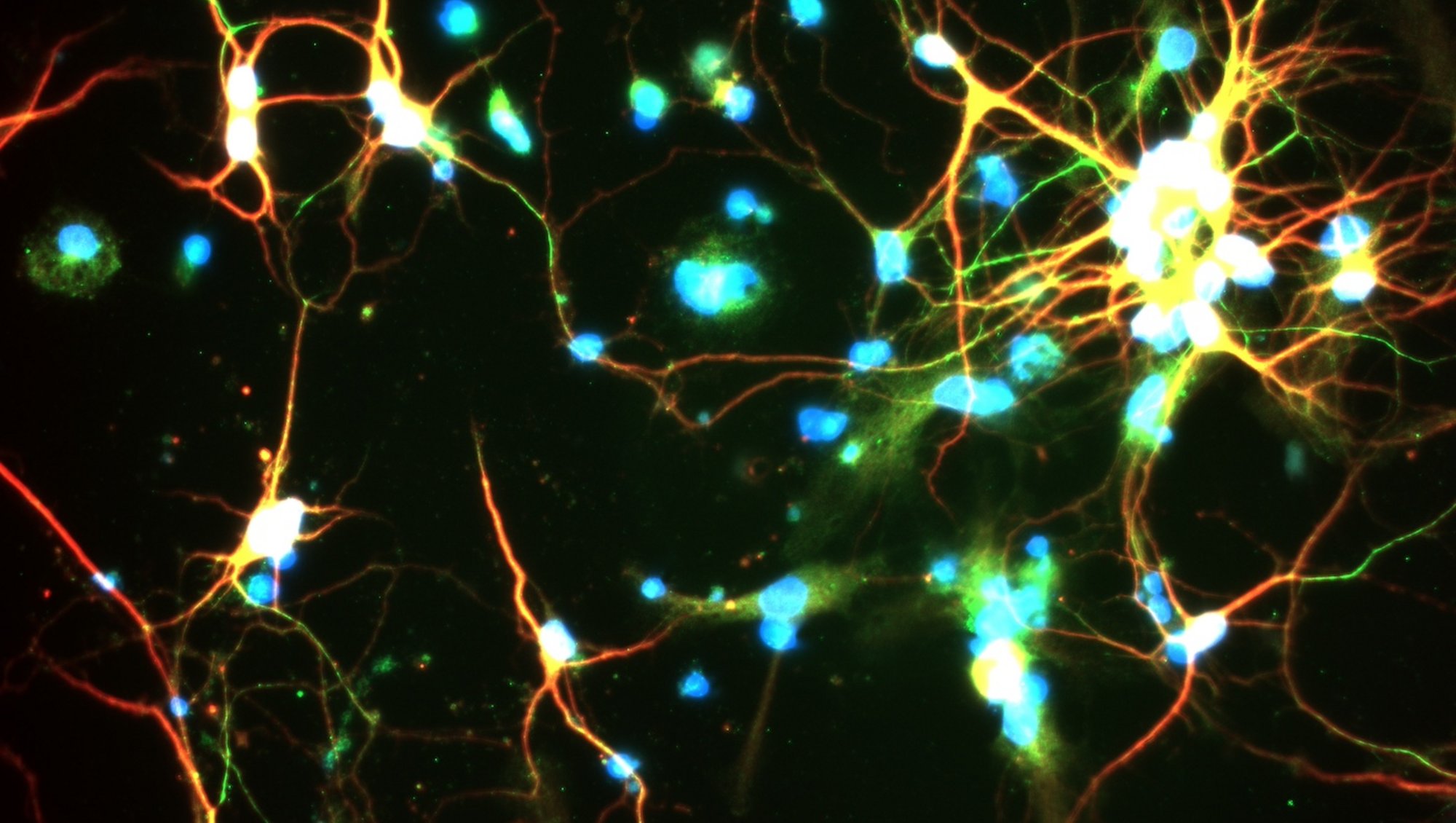
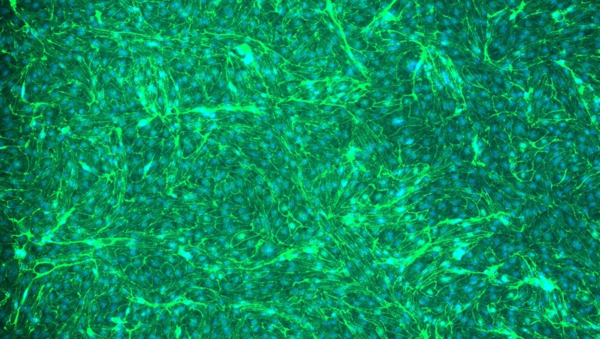
AxioObserver (Skou - 1116), Inverted Widefield, Zeiss
The AxioObserver (Skou) Inverted Widefield Microscope is easy to use while maintaining sensitivity.
Applications in brief
This inverted widefield microscope is equipped with two light sources and two corresponding detectors (cameras), allowing generation of either fluorescence images or brightfield imagages.
Its motorized stage allows automated sample finding and generation of tile scans.
In doubt if your staining worked? Book a 15 min time slot to check your sample.
Features
- Brightfield and/or multispectral fluorescence
- Stage mounting frames for slides (max. length 120 mm)
Which samples may I use?
Selected samples, but not limited to:
- fixed tissue sections or fixed cells
- brightfield (e.g. H&E)
- immunofluorescence (due to limtations by objectives, consider corresponding microscope located in the anatomy building for this application)
- live cells in thoroughly sealed containers
Training and use
Tips and tricks
Consider bleed-through/spectral overlap when investigating potential co-localization
- Ensure that the chosen fluorophores do not spectrally overlap.
- The current filtercubes of this microscope pose limitations to co-localization studies. Ensure that the settings (choice of filters ect.) are set correctly to avoid false co-localization signals or consider using the corresponding microscope located in the anatomy building.
Imaging of live cells
- For imaging of live cells, e.g. in petri dishes, ensure to seal the container with parafilm and to use a suitable stage to avoid spillage into the microscope and to ensure a horizontal sample.
Specifications
| Full equipment name | Widefield Inverted Light Microscope, Zeiss |
| Objectives |
|
| Filter qubes |
|
| Light sources |
|
| Detectors (cameras) |
|
| Heated stage | No |
| Motorized stage | Yes (tile scan possible) |


