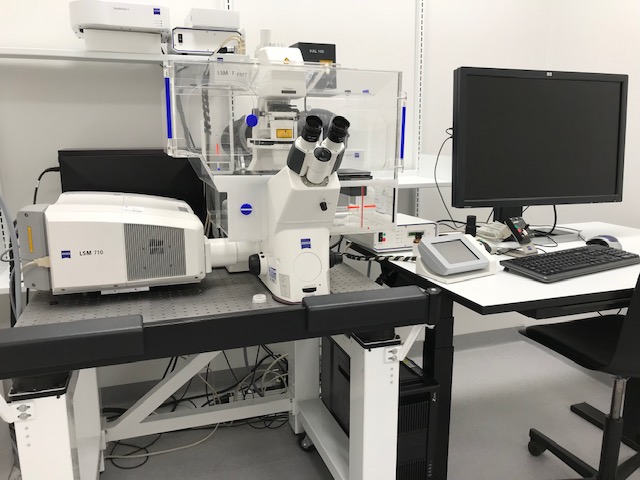
LSM 710, Laser Scanning Confocal, Zeiss


Would you like your image to be displayed here?


Please contact us or send an email to imaging@biomed.au.dk, if you would like to share your images - Thank you


The LSM 710 inverted laser scanning confocal microscope from Zeiss allows optical Z-sectioning of various fluorophores in living specimens and fixed samples.

Applications in brief

The LSM 710 can be used for a wide array of imaging applications on both live and fixed samples including multicolor imaging, protein co-localization, 3D z-stacks, and stitching as well as for time-lapse analysis for following cellular events in live samples.

Features

  • Motorized XY-stage
  • Temperature control
  • 3 detectors
  • 1 transmitted detector

Which samples may I use?

Selected samples, but not limited to:

  • living specimens
  • fixed cells
  • fixed tissue

Specifications

Full equipment name LSM710 inverted laser scanning confocal microscope, Zeiss
Objective Mag. / NA Medium Contrast Working Distance
Pln-Apo 20x/0.8 Air - 0,55
C Apochromat  40x/1,2 Water
DIC III  
0.28 at cover glass 0.17
Plan-Apochromat 63x/1.4 Oil DIC III 0.19
Lasers           Type and Power  Selected dyes                       
405nm Diode, 30mW DAPI, Hoechst 33342
458nm Argon, 25mW CFP
488nm Argon, 25mW GFP, AlexaFluor488
514nm Argon, 25mW YFP
543 nm HeNe, 5mW RFP
633nm HeNe, 5mW Cy5, AlexaFluor648

FAQ

Equipment

- How should I clean the equipment after use?

  • Please leave the work station neat and clean for the next user.
  • Please ensure that you have removed any oil from the oil objective.
  • In case of accidental contamination of air/water objectives with oil, please contact the bioimaging core facility staff immediately.

- Are consumables included in the price?

  • Experimental consumables are not included in the price for booking of microscopes via the Bioimaging Core Facility.
  • Designated oil for the oil objectives can be found next to the microscope. Please only use this designated oil for the objectives.
  • Lens paper designated for cleaning of objects as well as designated tissues for cleaning of samples are located next to the microscope.

Data Storage

- How can I get access to my data?

  • After using a microscope or a workstation, please transfer your data immediately to your personal AU drive (O:) and clean up your local directories on instrument PCs if you are not reusing data in subsequent experiments.
  • The Bioimaging Core Facility staff will provide support to non-AU users for exporting data.
  • Use of USB memory sticks or external hard drives is for security reasons not allowed.

- Does the Bioimaging Core Facility regularly back up the user data?

The Bioimaging Core Faility is not responsible for your data. DATA ON LOCAL PC DRIVES WILL BE DELETED ON A REGULAR BASIS in order to keep the systems performing optimally.

Please ensure that you transfer your data to your personal AU drive (O:) after each session.

- May I use my personal USB memory stick or external hard drive for data storage?

No, any use of USB memory sticks or external hard drives is for security reasons not allowed. Usage of USB memory sticks may lead to suspension of the user license.

Data Analysis

- How may I process and analyse my data?

  • Obtained images may be processed and analyzed using freeware or a selection of software available on PCs in the Bioimaging Core Facility.

Publications

- How shall I refer to the equipment?

Need help?

Come to our Open Office sessions to discuss your imaging needs or get advice on workflows, free of charge:

Tuesdays and Thursdays 13:30 -14:30

The Skou building, 1116-256