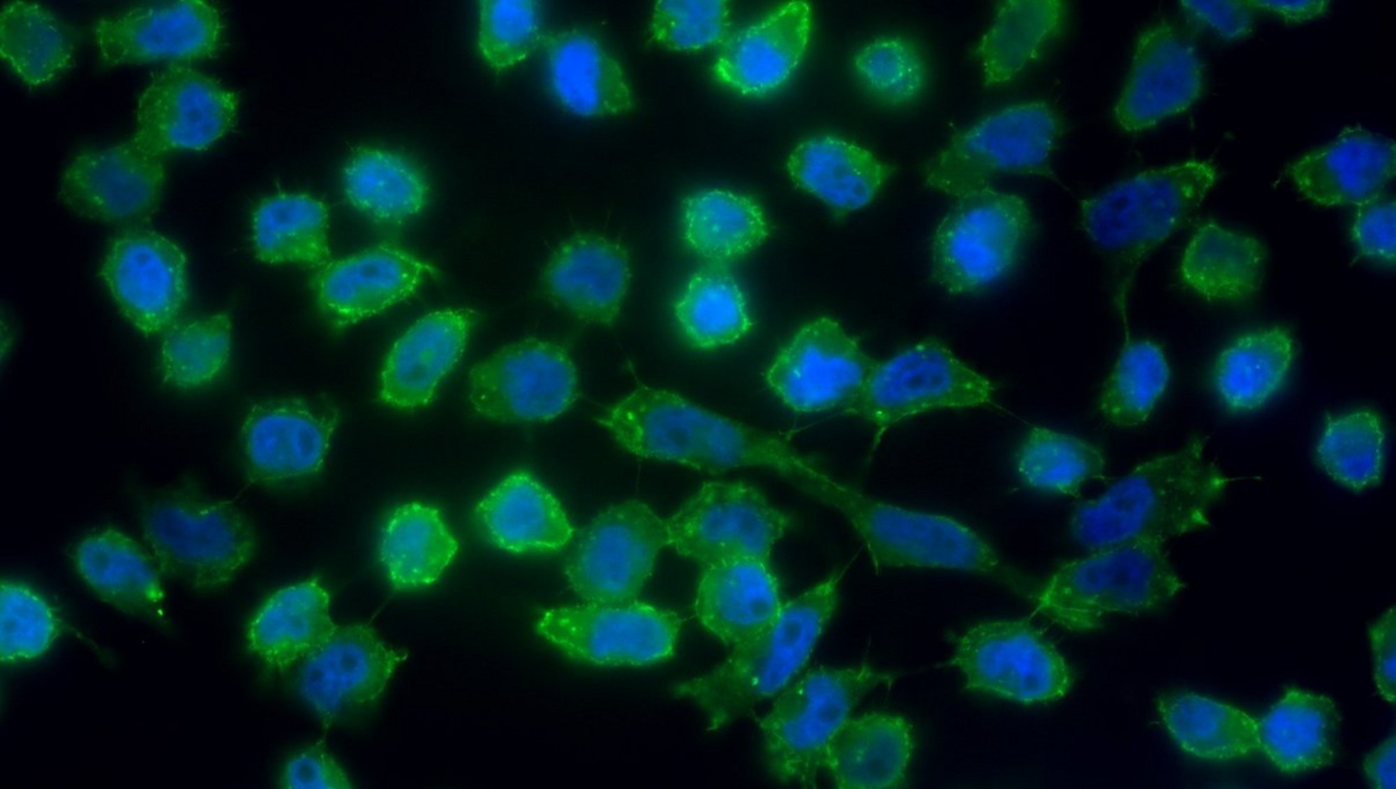
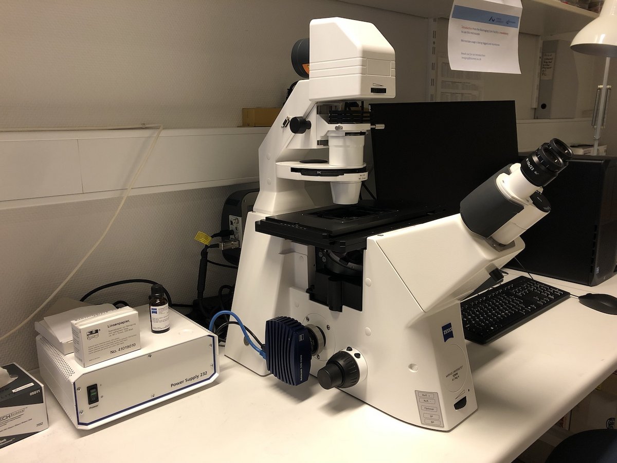
AxioObserver (Anatomy), Inverted Widefield, Zeiss

Would you like your image to be displayed here?

Please contact us or send an email to imaging@biomed.au.dk, if you would like to share your images - Thank you

The AxioObserver (Anatomy) Inverted Widefield holds a motorised stage and is easy to use while maintaining sensitivity.

Applications in brief

This inverted widefield microscope is equipped with two light sources and two corresponding detectors (cameras). This allows generation of either fluorescence images or brightfield imagages.

In doubt if your staining worked? Book a 15 min time slot to check your sample.

Features

  • Brightfield and/or multispectral fluorescence
  • Stage mounting frames for slides (max. length 120 mm) and petri dishes (Ø 24-68 mm)

Which samples may I use?

Selected samples, but not limited to:

  • fixed tissue sections or fixed cells
  • brightfield (e.g. H&E)
  • immunofluorescence

Price structure

Fixed Price Structure for Booking

The fixed annual price structure for AxioObserver Inverted Widefield usage allows unlimited booking of both the AxioObserver (Skou building) and the AxioObserver (Anatomy building). Within this structure, it is also possible for registered users to use the instruments for a quick sample check by booking a 15 min time slot.

Training Session

A training session is mandatory and is not included in the annual price. As the AxioObservers hold minor differences, the training session will be given to the most relevant AxioObserver and, if needed, supplemented with a free of charge, short add-on for the other AxioObserver.

Training and use

Tips and tricks


The image

  • Always locate and bring your sample in focus by looking through the occular before you play with software settings.

Specifications

Full equipment name Widefield Inverted Light Microscope, Zeiss
Objectives
  • 10x/0.25, Air, LongDistance A-Plan, coverslip thickness: 1 mm polystyrene
  • 40x/0.75, Air, EC Plan-Neofluar, coverslip thickness: 0.17 mm
  • 63x/1.40, Oil, Plan Apochromat, coverslip thickness: 0.17 mm
Filter qubes
  • Cy 5 (EX BP 640/30, BS FT 660, EM BP 690/50)
  • Cy3 (EX BP 550/25, BS FT 570, EM BP 605/70)
  • eGFP (EX BP 470/40, BS FT 495, EM BP 525/50)
  • Hoechst/DAPI (EX BP 390/40, BS FT 420, EM BP 450/40)
Light sources
  • Illuminator microLED (for brightfield applications)
  • Colibri 5 type RGB-UV (for fluorescence applications)
Detectors (cameras)
  • Axiocam 506 mono (for fluorescence applications)
  • Axiocam 105 color R2 (for brightfield applications) 
Heated stage No
Motorized stage No

FAQ

Equipment

- How should I clean the equipment after use?

  • If you have used the 63x Oil objective, ensure that you have gently wiped off the oil droplet with a piece of designated lens paper.
  • In case you accidentially contaminated air objectives with oil, please inform us immediately to ease the cleaning process for us as much as possible.
  • Please only use designated lens paper for wiping off oil from objects and designated tissues for cleaning of samples.
  • Please leave the work station neat and clean for the next user.
  • Ensure to cover the microscope with the designated dust cover.

- Are consumables included in the price?

  • Experimental consumables are not included in the price for booking of microscopes via the Bioimaging Core Facility.
  • Designated oil for the oil objectives can be found next to the microscope. Please only use this designated oil for the objectives.
  • Lens paper designated for cleaning of objects as well as designated tissues for cleaning of samples are located next to the microscope.

Data Storage

- How can I get access to my data?

  • After using a microscope or a workstation, please transfer your data immediately to your personal AU drive (O:) and clean up your local directories on instrument PCs if you are not reusing data in subsequent experiments.
  • The Bioimaging Core Facility staff will provide support to non-AU users for exporting data.
  • Use of USB memory sticks or external hard drives is for security reasons not allowed.

- Does the Bioimaging Core Facility regularly back up the user data?

The Bioimaging Core Faility is not responsible for your data. DATA ON LOCAL PC DRIVES WILL BE DELETED ON A REGULAR BASIS in order to keep the systems performing optimally.

Please ensure that you transfer your data to your personal AU drive (O:) after each session.

- May I use my personal USB memory stick or external hard drive for data storage?

No, any use of USB memory sticks or external hard drives is for security reasons not allowed. Usage of USB memory sticks may lead to suspension of the user license.

Data Analysis

- How may I process and analyse my data?

  • Obtained images may be processed and analyzed using freeware or a selection of software available on PCs in the Bioimaging Core Facility.

Publications

- How shall I refer to the equipment?

Need help?

Come to our Open Office sessions to discuss your imaging needs or get advice on workflows, free of charge:

Tuesdays and Thursdays 13:30 -14:30

The Skou building, 1116-256